
The Department of Veterinary Anatomy is one of the constituent departments of the Arawali Veterinary College since its inception in the year 2009. The mandate of the department is to impart education and practical training in courses of veterinary anatomy and histology to the students preparing for the degree of B.V.Sc.&A.H. Sound knowledge of body structure will be base for understanding physiological functions and identification of sites for performing operations on sick animals.
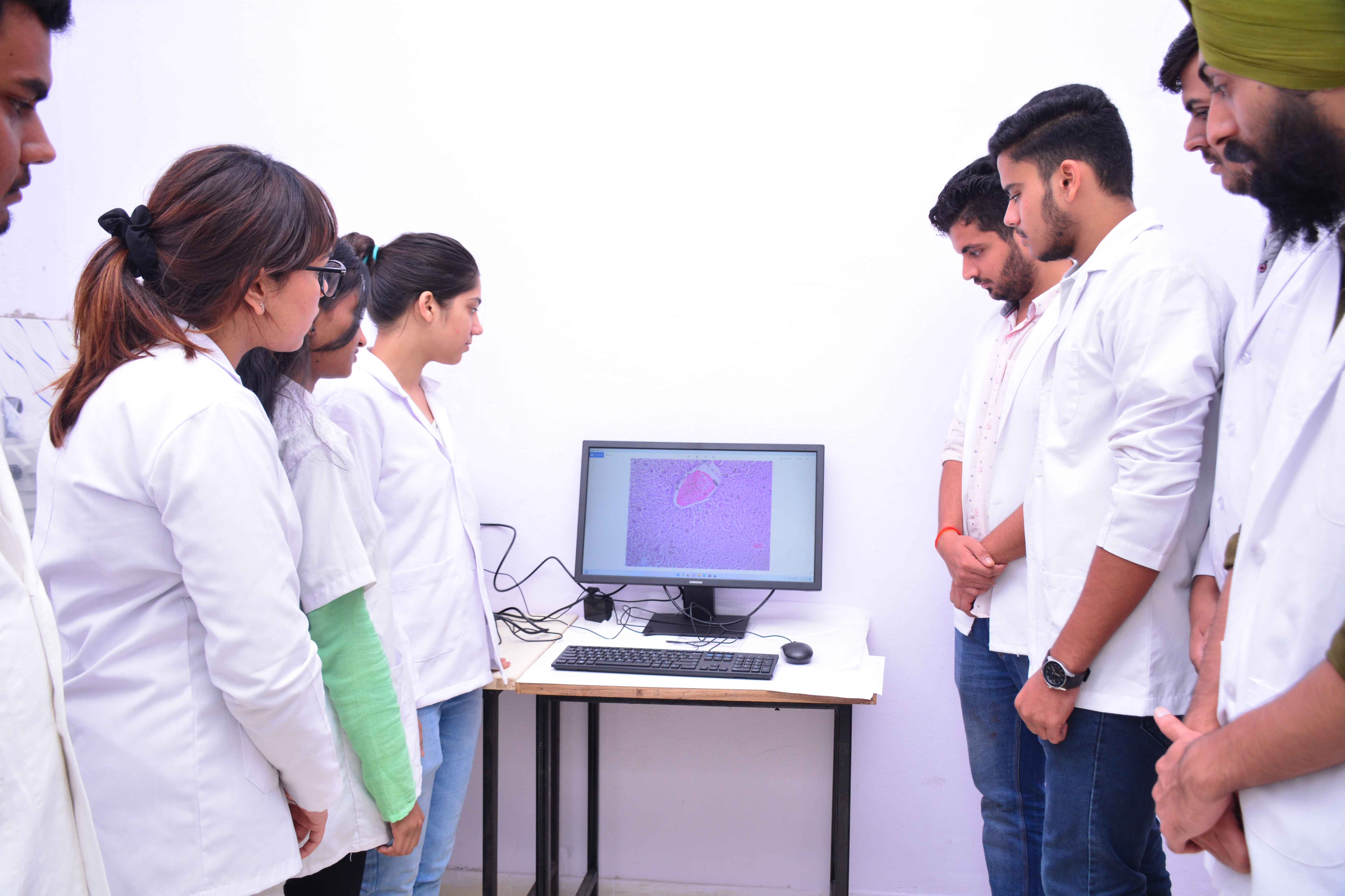
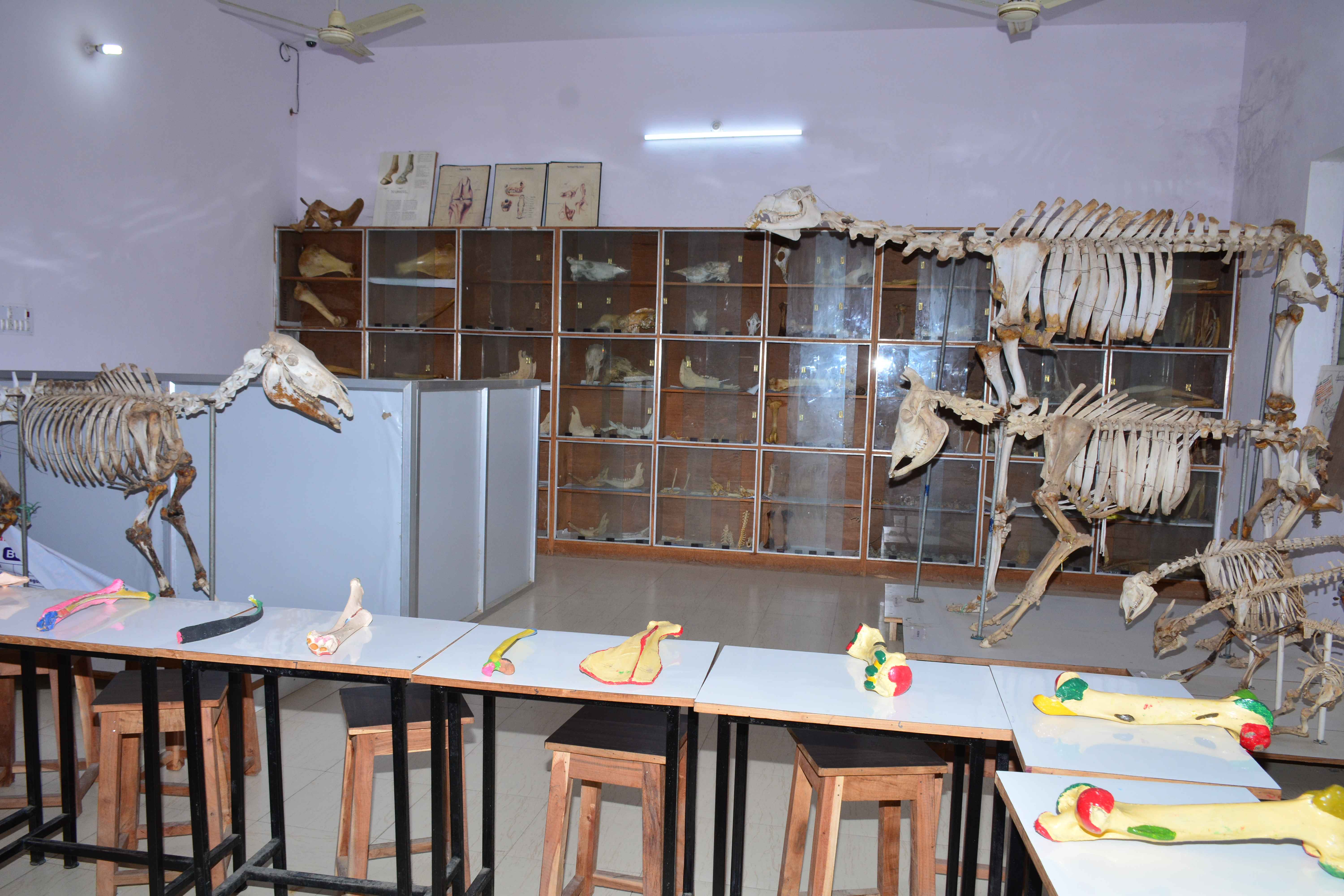
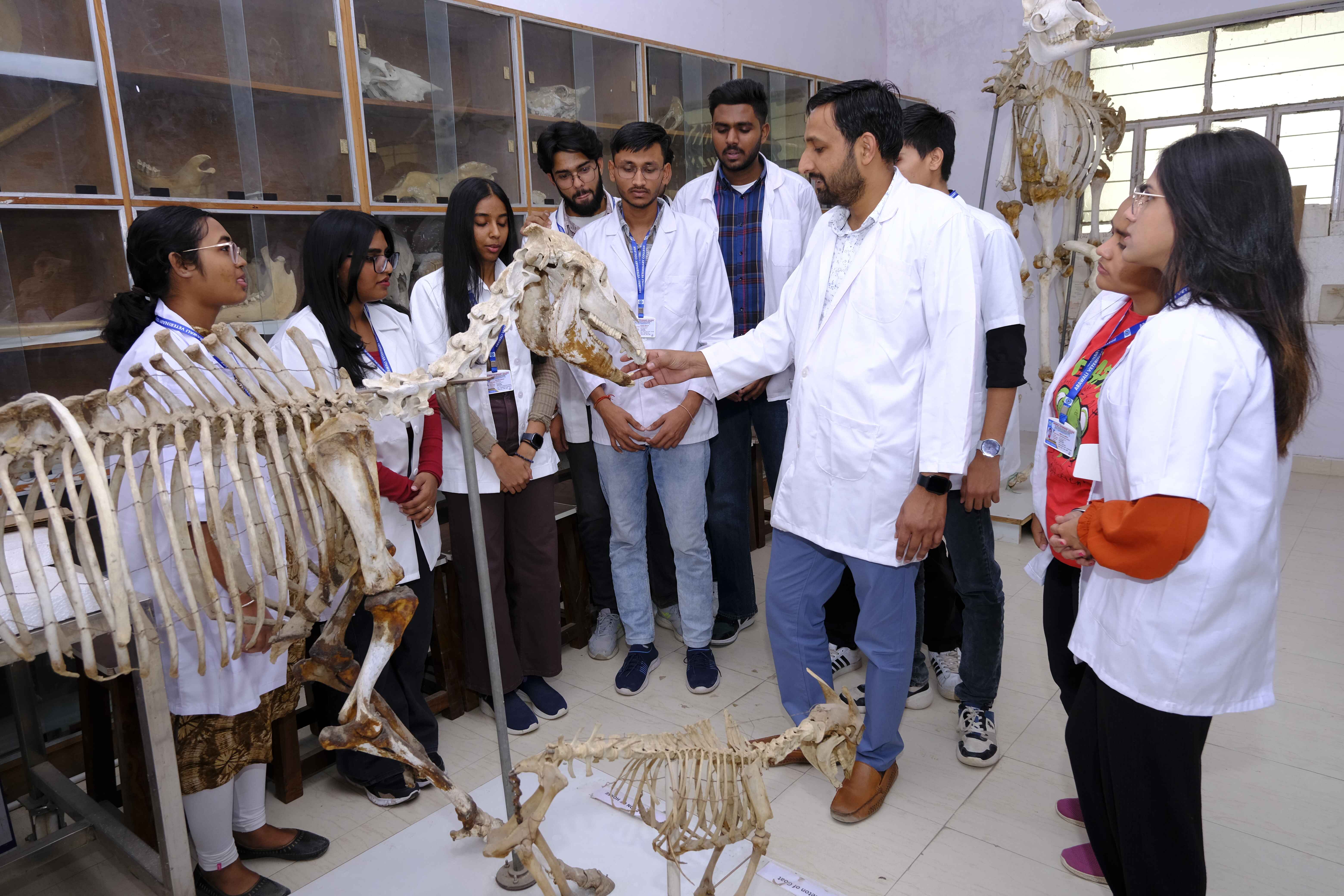
The department has a good collection of skeletons and visceral organs of all domestic, pet and some wild animals in its museum. Dissections are carried out on cadavers prepared from carcasses donated after the postmortem of dead animals. In addition, models, mannequins, plastinated specimens, and preserved body organs are used to impart practical training for a better understanding of the structure and functions of the animal body. Dissections are carried out on cadavers procured by way of donation of animal carcasses or obtained from postmortem section.Computer-simulated software, laboratory manuals and audio-visual facilities are used as tools for an in-depth understanding of the subject.
The department has well equipped laboratories for conducting the following activities.
- Dissection of animals' corpse.
- Embalming of dead animals.
- Preparation of microscopic slides for teaching histology.
- Procurement through donation, and processing of bones of various animals (limbs, Skull and vertebrae etc.)
The Department has the following sanctioned strength of Faculty:
Professor - 1 Associate Professor – 1 Assistant Professor – 2
|
S.NO
|
Name
|
Designation
|
Qualification
|
Service Period at AVC
|
Photo
|
Biodata
|
|
01
|
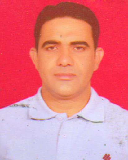
Dr Vikas Kumar
|
Assistant Professor
|
B.V.Sc & AH, M.V.Sc
|
27.01.2017 to till continue
|

|
Biodata
|
|
02
|
Dr. Sanwar Mal
|
Assistant Professor
|
B.V.Sc & AH, M.V.Sc
|
09.12.2022 to till continue
|
|
Biodata
|
|
03
|
Dr. Pawan Kumar
|
Assistant Professor
|
B.V.Sc & AH, M.V.Sc
|
02.04.2025 to till continue
|
|
Biodata
|
SYLLABUS
THEORY
UNIT:1
Introduction to anatomy and branches of anatomy and descriptive terms used in anatomy and study of anatomical planes. General Osteology, Arthrology and Myology: Study of properties and structure of bone. Classification of skeletons, classification of bones with suitable examples and terms used in osteology Introduction to arthrology, classification of joints, different diarthrodial joints, structure of diarthrodial joints and movements permitted. Introduction to myology, classification of muscles, etymology of muscles. Description of tendon, ligaments, aponeurosis, synovial bursa and synovial sheath.
(Note: Detailed description of muscles of different regions of the body will be studied in the respective practical).
General Angiology, Neurology and Aesthesiology: Introduction to angiology. Structure of heart. General plan of systemic and pulmonary circulations, lymphatic and venous systems. Introduction to neurology and parts of central, peripheral and autonomic nervous system and sense organs. Formation of spinal nerve. Structure of meninges, brain, spinal cord. Different surface regions, joint regions, Palpable Bony areas or prominences of the body of the animal. Palpable Lymph nodes and Arteries of the body and Surface veins for Venepuncture. Sites for collection of Bone marrow and Cerebrospinal fluid. General Splanchnology: Introduction to splanchnology, boundaries of thoracic, abdominal and pelvic cavities, topography of different organs of digestive, respiratory, urinary, endocrine, male and female reproductive systems of domestic animals and fowl. Principles and application of Radiography and Ultrasound for bones and soft tissues.
UNIT-2
Fore limb: Study of bones of fore limb of ox and differences in horse, dog, pig and fowl. Study of hoof of ox and horse. Study of joints, ligaments, stay apparatus, major blood vessels, nerves, veins and lymph nodes of fore limb. Sites for Radial, Median, Ulnar and Volar nerve blocks.
UNIT-3
Head and neck: Study of cranial and facial bones, cervical vertebrae of ox and differences in horse, dog, pig and fowl. Boundaries of the oral, orbital, nasal and cranial cavities. Study of paranasal sinuses in ox, horse, dog and pig. Study of articulations and special ligaments of the head and neck. Muscles of face, mastication, eye, ear, tongue, pharynx, soft palate,hyoid and larynx. Study of teeth, hard and soft palate, tongue, pharynx, larynx, thyroid, parathyroid and salivary glands and differences in horse, dog, pig and fowl. Study of cranial nerves, blood vessels and lymph nodes of head and neck regions. Study of boundaries of jugular furrow and structures of carotid sheath along with neck muscles. Study of sense organs, trachea and oesophagus. Age determination by Dentition. Sites for Tracheotomy, Esophagotomy, Ligation of Stensons duct and Mental, Mandibular, Maxillary , Cornual, Infraorabital , Supraorbital (frontal) , Orbital and Auriculopalpebral nerve blocks and surgical approach to guttural pouches in horse. Importance of Cornual nerve and superficial temporal artery in Amputation of Horn in cattle.
UNIT-4
Thorax: Study of thoracic vertebrae, ribs and sternum of ox and differences in horse, dog, pig and fowl. Study of joints, special ligaments, blood vessels, nerves, lymph vessels and lymph nodes of thorax. Study of organs of thorax i.e. trachea, thymus, oesophagus, lungs and differences in horse, dog, pig and fowl. Study of pleura, its reflections and mediastinum. Areas of auscultation and percussion of heart and lungs and site for Paracentesis Thoracis.
UNIT-5
Abdomen: Study of bones of abdomen of ox and differences in horse, dog, pig and fowl. Study of joints, special ligaments blood vessels, nerves of abdomen region. Blood and nerve supply to abdominal viscera. Study of peritoneal reflections, organs of digestive, urinary, male and female reproductive systems present in abdomen and differences in horse, dog, pig and fowl. Study of mammary glands in cow and differences in mare, bitch and sow. Study of spleen of ox and differences in horse, dog, pig and fowl. Study of major veins, lymph vessels, lymph nodes and endocrine glands of abdomen. Boundaries and Clinical importance of the flank and Para Lumbar Fossa. Sites for Liver ,Gall Bladder and Caecal Biopsies, Laparotomy,Rumenocentesis Operation , enterotomy, and paravertebral block .
UNIT-6
Hind limb and pelvis: Study of bones of hind limb and pelvis of ox and differences in horse, dog, pig and fowl. Study of joints, ligaments, blood vessels, lymph nodes and nerves of hind limb, pelvis and tail region and pelvic viscera. Study of pelvic peritoneal reflections, organs of digestive, urinary, male and female reproductive systems present in pelvic cavity and differences in horse, dog, pig and fowl. Boundaries of the inguinal canal and structures of the spermatic cord, pre pubic tendon and its importance. Study of external genital organs. Sites for Tibial , Peroneal ,Plantar and Pubic nerve blocks, Patellar desmotomy, Urethrotomy, Castration , Vasectomy, cranial and caudal epidural anaesthesia.
UNIT-7
Cytology, cell junctions, study of basic tissues i.e epithelial, connective, muscular and nervous tissues, blood and bone marrow. Study of microscopic structures of digestive, circulatory, urinary, respiratory, nervous, lymphatic, endocrine, male and female genital systems and mammary glands of domestic animals. Study of microscopic structure of sense organs i.e. eye, ear and integument.
UNIT-8
Introduction to embryology, gametogenesis, fertilization, cleavage, types of eggs, morula, blastulation, gastrulation, types of implantation, twinning. Formation of foetal membranes in mammals and birds, Placenta and its classification. Different germ layers and their derivatives. Study of development of organs of digestive system including accessory structures i.e tongue, teeth, salivary glands, liver and pancreas. Study of development of organs of respiratory, urinary, circulatory, lymphatic, nervous, musculoskeletal, male and female reproductive systems. Development of endocrine glands, sense organs i.e eye and ear.
PRACTICAL
UNIT-1
Study of general terms used in anatomy, study of anatomical planes. Study of different parts of skeleton, different surface and joint regions. Study of boundaries of thoracic, abdominal and pelvic cavities. Demonstration of different types of joints, muscles tendons, ligaments, synovial bursa and synovial sheath. In situ demonstration of heart, meninges, brain and spinal cord. Boundaries of Thoracic, Abdominal and Pelvic Cavities and in situ demonstration of organs of digestive, respiratory, urinary, endocrine, male and female reproductive systems of domestic animals. Demonstration of Different surface regions, joint regions and Palpable Bony areas or prominences of the body of the animal , Common sites of fractures, Palpable Lymph nodes and Arteries of the body ( ventral coccygeal artery in ox, femoral artery in dog and cat , facial artery in horse ) and Surface veins for Venepuncture ( cephalic vein and recurrent tarsal vein in dog and cat , jugular vein in large animals.) and Sites for collection of Bone marrow and Cerebrospinal fluid. Visualization of Radiographs and ultrasound pictures of various organs and Fractures of various bones.
UNIT-2
Fore limb: Demonstration of different bones of fore limb of ox and comparison with horse, dog, pig and fowl. Dissection of the fore limb. Study of joints, ligaments, muscles, major blood vessels, lymph nodes and nerves of fore limb. Study of sites for different nerves blocksorneurectomies in fore-limb. Study of suprascapular nerve paralysis shoulder sweeny, radial nerve paralysis-capped elbow. Structure of the equine hoof and comparison with ox. Demonstration of radiographs of normal bones of fore limb. Clinical importance of cephalic vein for intravenous injections in dog.
UNIT-3
Head and neck: Demonstration of cranial and facial bones, cervical vertebrae of ox and comparison with horse, dog and fowl. Dissection of muscles of face, mastication, tongue, pharynx, soft palate, hyoid, larynx, eye and ear. Dissection of superficial neck muscles. Dissection of brain and its parts. Dissection or demonstration of tunics of eye. Study of teeth, tongue, pharynx, thyroid, parathyroid and salivary glands and differences in horse, dog, pig and fowl. Study of cranial nerves, and blood vessels of head and neck regions. Study of trachea and oesophagus. Study of nerve blocks of the head i.e cornual, auriculo-palpebral, Peterson’s orbital nerve block, mandibulo-alveolar and mental nerve blocks. Importance of facial artery for recording pulse in horse. Surgical importance of Stenson’s duct in domestic animals. Surgical approach to guttural pouches-Viborg’s triangle. Clinical importance of jugular vein for intravenous injections in large animals. Demonstration of radiographs of normal bones of head and neck.
UNIT-4
Thorax: Demonstration of thoracic vertebrae, ribs and sternum of ox and comparison with horse, dog, pig and fowl. Dissection of muscles, blood vessels, nerves and lymph nodes of thorax. Demonstration of organs of thorax i.e. trachea,oesophagus, thymus, lungs and heart and differences in horse, dog, pig and fowl. Study of pleural reflections of thoracic cavity. Demonstration of sites for auscultation and percussion. Recurrent laryngeal nerve paralysis-roaring in horses. Choke or oesophageal obstruction. Demonstration of radiographs and videos of ultrasonography of organs of thorax.
UNIT-5
Abdomen: Demonstration of bones forming boundaries of abdomen of ox and comparison with horse, dog, pig and fowl. Dissection of muscles, blood vessels and nerves of abdomen. Demonstration of peritoneum, omentum, mesentry and organs of digestive, urinary, male and female reproductive systems present in abdomen and differences in horse, dog, pig and fowl. Demonstration of mammary glands of cow, mare, bitch and sow. Demonstration of major veins, lymph vessels and lymph nodes of abdomen. Topographic location of abdominal viscera of ox and comparison with horse, dog, pig and fowl. Demonstration of sites for laparotomy, caesarean section, ovario-hysterectomy, catheterization of urinary bladder and sites for paravertebral and epidural anaesthesia. Demonstration of Boundaries and Clinical importance of the flank and Para Lumbar Fossa, Sites for Liver ,Gall Bladder and Caecal Biopsies, Laparotomy, Rumenocentesis, Rumenotomy, abomasotomy, spleenectomy Cystotomy, Caesarean Operation, catheterization of urinary bladder and enterotomy and paravertebral block . Demonstration of radiographs and videos of ultrasonography of organs of abdomen.
UNIT-6
Hind limb and pelvis: Demonstration of bones of hind limb of ox and comparison with horse, dog, pig and fowl. Demonstration of joints and ligaments of hind limb and pelvis. Dissection of muscles, blood vessels, lymph nodes and nerves of hind limb and pelvic cavity. Demonstration of peritoneal reflections of pelvic cavity and organs of digestive, urinary, male and female reproductive systems in pelvic cavity and differences in horse, dog, pig and fowl. Study of external genital organs. Clinical importance of femoral artery to record pulse in dog. Clinical importance of recurrent tarsal vein for intravenous injections in dog. Demonstration of radiographs of normal bones and videos of ultrasonography of organs of pelvis. Demonstration of Sites for Tibial, Peroneal ,Plantar and Pudic nerve blocks, Patellar desmotomy, Urethrotomy, Castration , Vasectomy and cranial and caudal epidural anaesthesia.
UNIT-7
Microscopy and micrometry. Comparison of light and electron microscopy. Histological techniques, processing of tissues for paraffin sectioning and haematoxylin and eosin staining. Microscopic examination of epithelium, connective tissue, muscular tissue, nervous tissue and blood. Microscopic examination of organs of digestive, circulatory, urinary, respiratory, nervous, lymphatic, endocrine, male and female genital systems and sensory organs of domestic animals.
UNIT-8
Demonstration of Placenta, umbilical cord and foetal membranes of different domestic animals. Demonstration of congenital anomalies of domestic animals as per availability. Study of slides of developing organs of different systems as per the availability. A embalmed cadaver of buffalo calf (procured through donated animals or cadevars obtained from post-mortem section) for every 24 students to be used for dissection purposes.
ANNUAL EXAMINATION
PAPERS UNITS MAXIMUM MARKS WEIGHTAGE
THEORY
Paper-I, 1, 2, 3and 4 100 20
Paper-II 5,6,7 and 8 100 20
PRACTICAL
Paper-I 1, 2, 3and 4 60 20
Paper II 5,6,7 and 8 60 20
20